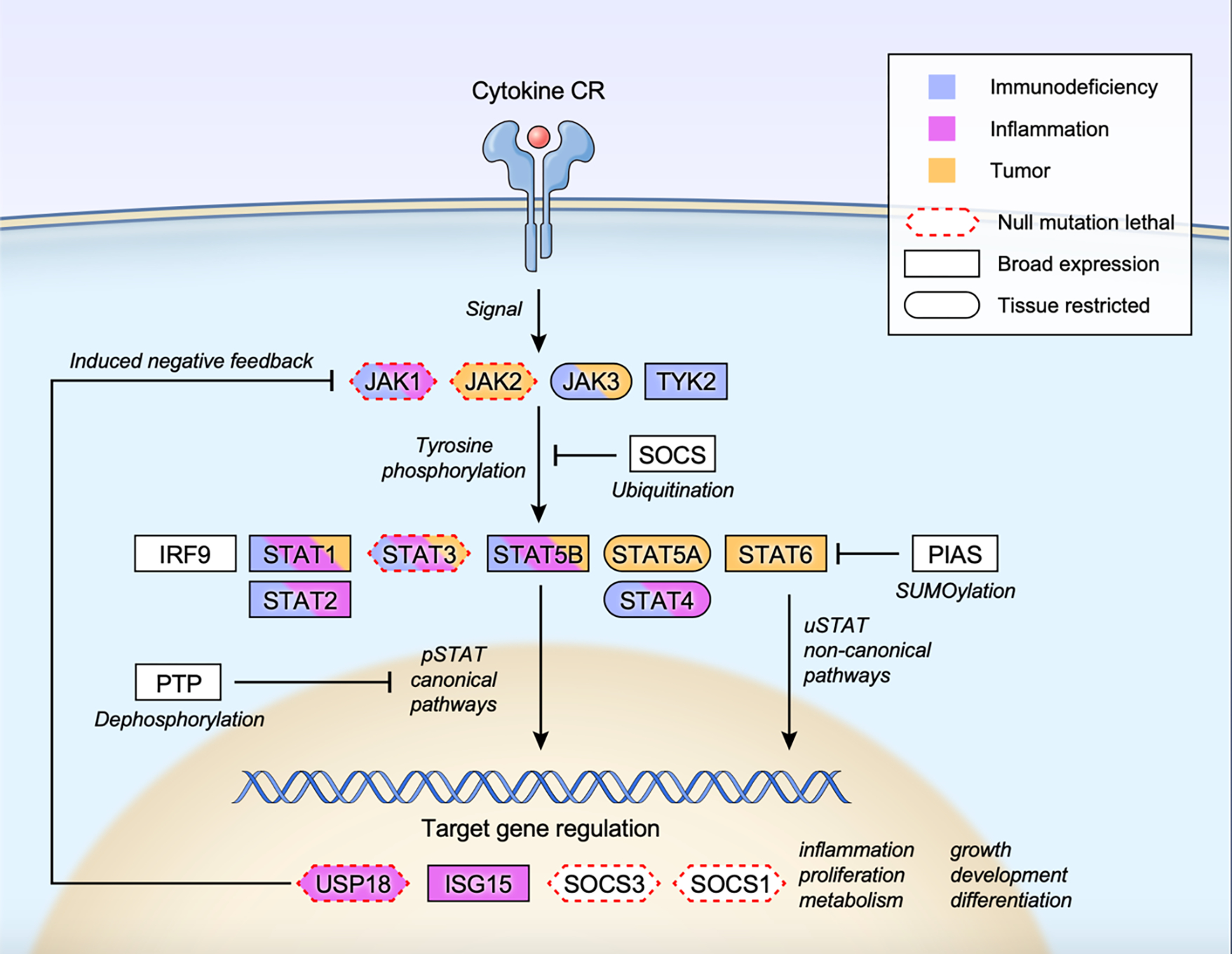
Figure 2. Overall scheme of JAK STAT signaling.

Cytokine-receptor engagement transmits signal to tyrosine phosphorylate and activate receptor associated JAKs, which subsequently phosphorylate and activate STATs. Tyrosine phosphorylated STATs (pSTATs) form dimers, translocate to the nucleus, bind to target DNA sequences, and regulate gene expression (pSTAT canonical pathways). Unphosphorylated STATs also form dimers, enter the nucleus and regulate transcription (uSTAT non-canonical pathways). Multiple inducible negative feedback systems are in place to constrain the signaling circuit that include SOCS (suppressor of cytokine signaling) family proteins, PIAS (protein inhibitor of activated STAT) family proteins, PTP (protein tyrosine phosphatase), USP18 and ISG15. Human monogenic mutations that lead to gain-of-function and/or loss-of-function phenotypes are color coded as immunodeficiency (blue), inflammation (pink) or tumor (orange) for each constituent gene. Genes whose null mutation in mice leads to lethality is marked with red dotted border (Mouse Genome Informatics; http://www.informatics.jax.org). Tissue expression pattern of each protein is also marked as broad expression (square border) or tissue restricted (oval border) (The human protein atlas; https://www.proteinatlas.org/search).
