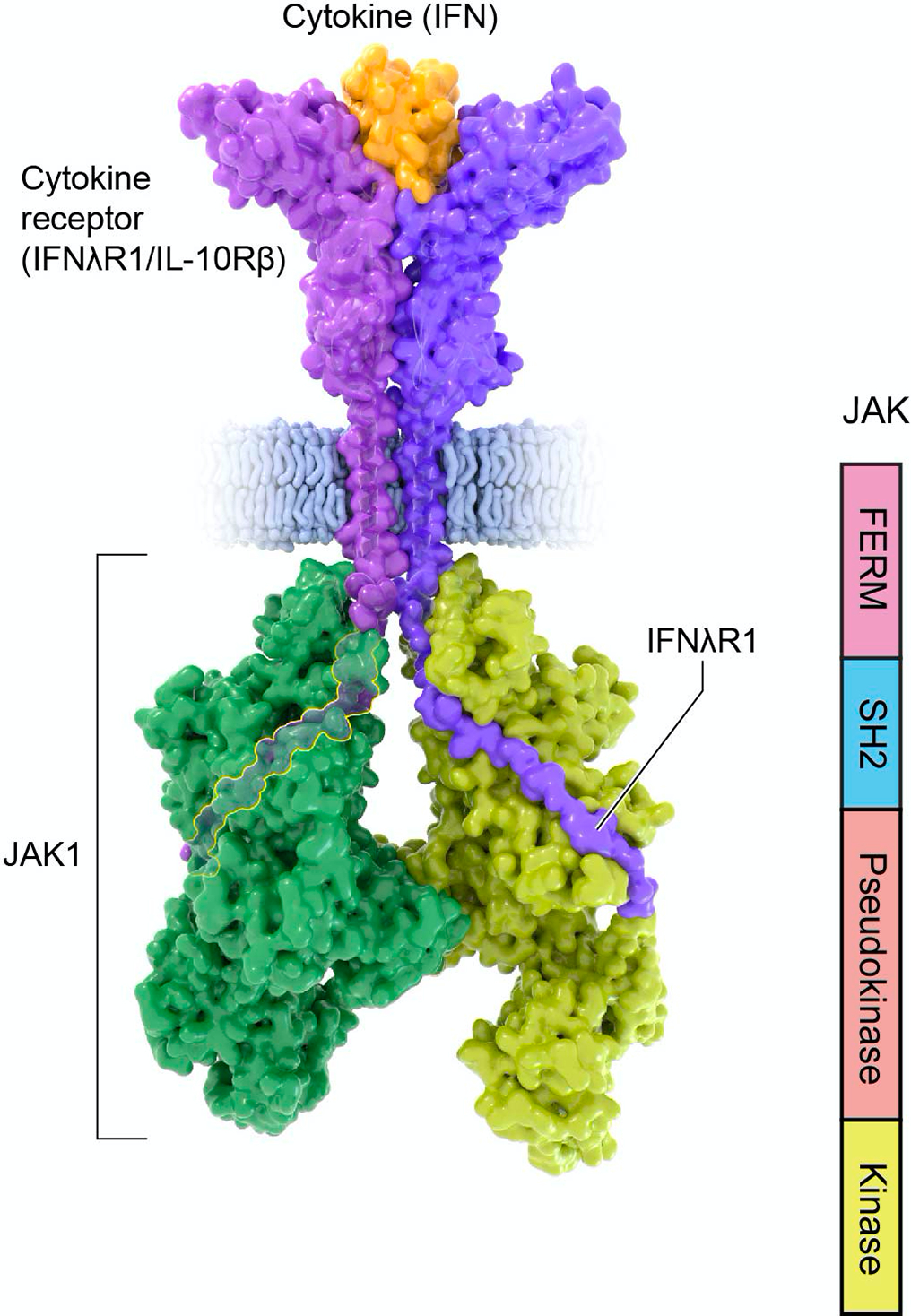
Figure 3. Structure of Janus kinases.

Structure of activated Janus Kinase dimer (green; PDB 7T6F) complexed with the intracellular domain of matured IFNlR1/IL-10Rb (purple) bound to IFN-l (orange) (PDB 5T5W). Of note, the intracellular portion of the receptor binds JAK1 FERM and SH2 domains through N-terminal Box1 and C-terminal Box2 motifs. The JAK kinase-like or pseudokinase domain promotes dimerization of the cytokine receptor/JAK complex.
