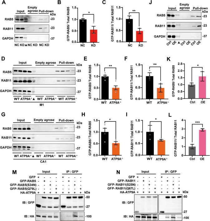
Fig. 6. ATP9A promotes the activation of RAB5 and RAB11.
A–C The active form of RAB5 and RAB11 (GTP-RAB5 and GTP-RAB11) in control (NC) and ATP9A knockdown (KD) N2a cells was determined by GTP-agarose pull-down assay. A Representative immunoblots of total or active RAB5 and RAB11 in cells were detected by western blots. “Empty agarose” indicates that samples were incubated with blank agarose beads, and “Pull-down” indicates that samples were incubated with GTP-agarose beads. B, C Quantification of RAB5 and RAB11 activity in control and ATP9A knockdown N2a cells (n = 3). GTP-RAB5 and GTP-RAB11 in M1 (D–F) and CA1 regions in the hippocampus (G-I) of WT and ATP9A−/− mice were determined by GTP-agarose pull-down assay. Representative immunoblots of total or active RAB5 and RAB11 in M1 (D) and hippocampal CA1 (G) regions of WT and ATP9A−/− mouse brains were detected by Western blots. (E, F, H, I) Quantification of RAB5 and RAB11 activity in M1 (n = 5) and hippocampal CA1 (n = 4) regions of WT and ATP9A−/− mouse brains. J–L GTP-RAB5 and GTP-RAB11 were determined by GTP-agarose pull-down assay in control (Ctrl) and ATP9A overexpressed (OE) N2a cells (n = 3). M HA-ATP9A were co-transfected with GFP-vector, GFP-RAB5 (Q79L) or GFP-RAB5 (S34N) in N2a cells. GFP-tagged RAB5 was immunoprecipitated from cell lysates and immunoblotted against exogenous ATP9A. N HA-ATP9A was co-transfected with GFP-vector, GFP-RAB11 (Q70L), or GFP-RAB11 (S25N) in N2a cells. GFP-tagged RAB11 was immunoprecipitated from cell lysates and immunoblotted against exogenous ATP9A. All values are presented as mean ± SEM (*P < 0.05, **P < 0.01, and ***P < 0.001, unpaired t-test for B, C, E, F, H, I, K and L). Filled triangles indicate protein markers.

