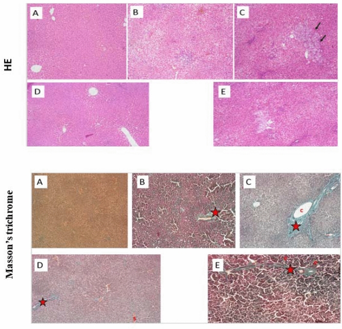
Figure 3.
The effect of early and late Kefir treatment on hepatic steatosis, inflammation and fibrosis using HE and Masson’s trichrome staining (Magnifications: × 100). (A) Control group showed no pathological changes, (B) NASH (9 weeks) group developed mainly micro vesicular steatosis, mild hepatic lobular inflammation and mild fibrous septa(star) (C) NASH (12 weeks) group showed diffuse and extensive micro and macro-vesicular steatosis, severe lobular inflammation, focal necrosis, hepatocellular ballooning, marked expansion of facultative hepatic progenitor cells (arrows), dense fibrous septa and collagen fibers (star) (D) Early Kefir and (E) Late Kefir groups showed a lower degree of steatosis, inflammation and fibrosis, where Early Kefir showed more remarkable histological improvements.

