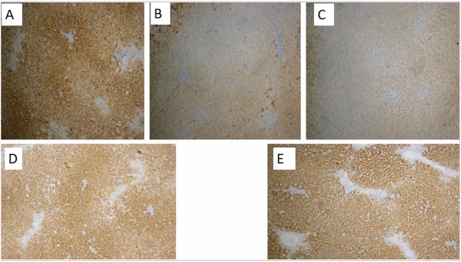
Figure 5.
Immunohistochemical staining for hepatocyte specific antigen (HepPar1). (A) Controls showed a strong signal with Hep Par 1 (hepatocyte marker) producing distinct granular, cytoplasmic staining of hepatocytes, whereas in both (B) NASH (9 weeks) and (C) NASH (12 weeks) groups were mostly negative with HepPar-1. By comparison, (96%) hepatocytes in (D) Early Kefir group were immunoreactive for HepPar-1 and (E) Late Kefir group tended to show weaker, patchy positivity (HepPar1 × 100) indicated to hepatocyte differentiation.

