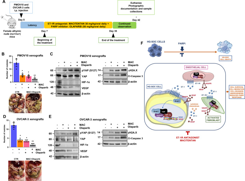
Fig. 8. Macitentan reduces HG-SOC metastatic potential and sensitizes to olaparib in vivo.
A Treatment schedule of patient-derived HG-SOC xenografts (PDX) and OVCAR-3 xenografts. B, D The number of tumor nodules examined at the end of the treatment. Bars are the means ± SD (*p < 0.0002 vs. vehicle-treated mice (CTR); **p < 0.0004 vs. olaparib-treated mice; n = 2). Right panels, Representative i.p. The metastatic nodules are indicated by white dotted-line circles. C, E pYAP (S127), YAP, HIF-1α, VEGF, γH2A.X and cl-caspase 3 protein expression in total cell lysates of i.p. nodules was evaluated by IB analysis. β-actin represents the loading control. Representative images of blots of 2 independent experiments are shown. F Working model illustrating how under the guidance of the ET-1/ET-1R axis, mutp53 anchors YAP and HIF-1α to DNA, turning on a cooperative transcriptional program in HG-SOC cells, endothelial cells and activated fibroblasts that culminates with the release of soluble mediators, such as ET-1, and how the amplification of a self-feeding circuit blunts the effect of PARPi. ET-1R blockade, dismantling the cross-talk between HG-SOC, EC, and activated fibroblasts, empowers olaparib efficacy, representing a valid companion for PARPi-based therapy.

