Fig. 4.
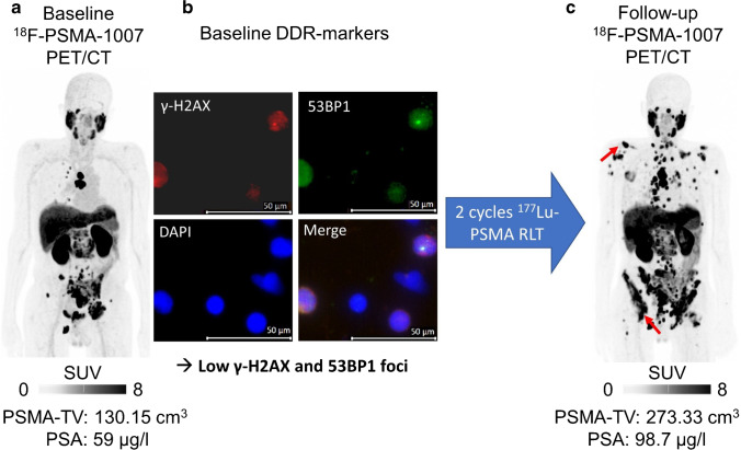
Low DDR-marker levels are associated with treatment failure. Baseline [18F]F-PSMA-1007 PET/CT (maximum intensity projection (MIP), a) of a patient prior to treatment. This patient had multiple tumor lesions in lymph nodes and the skeleton, e.g., in the11th thoracic vertebral body. Averaged SUVmax was 18.39 (above median, indicative of no progressive disease). Pretherapeutic PSA was 59 µg/l. Fluorescence microscopy images (63 × objective lens at 1.6 × magnification), with one merged and three separate channels for γ-H2AX (red), 53BP1 (green), and DAPI for nuclei stain (blue) of DDR-markers in PBLs prior to treatment (b). This patient demonstrated a low number of γ-H2AX (n = 0.16) and 53BP1 (n = 0.18) foci per cell, presumably reflecting low radiosensitivity. This patient had progressive disease after 2 cycles of [177Lu]Lu-PSMA RLT (c; PET: 71 days after study inclusion, multiple new lesions (red arrows) on follow-up PET/CT; PSA: 65 days after study inclusion, + 67% compared to baseline)

