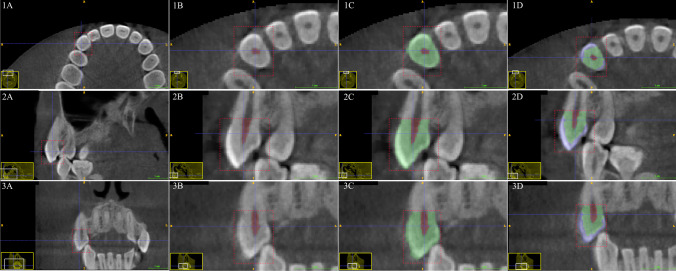
Fig. 1.
Segmentation sequence in the ITK-SNAP interface for the maxillary canine. Segmentation sequence as seen in column 1 (A–D) = axial view, column 2 (A–D) = sagittal view, column 3 (A–D) = coronal view. Row A = initial region of interest placement (red box), row B = pulp chamber volume label (red), row C = whole crown segmentation (green), row D = separation between dentine volume (green) and enamel volume (blue). The yellow window (bottom left) visualizes the current navigation region from the overall CBCT scan section plane with white inset box for the enlarged image. The blue cross represents the target point of interest for CBCT scan navigation

