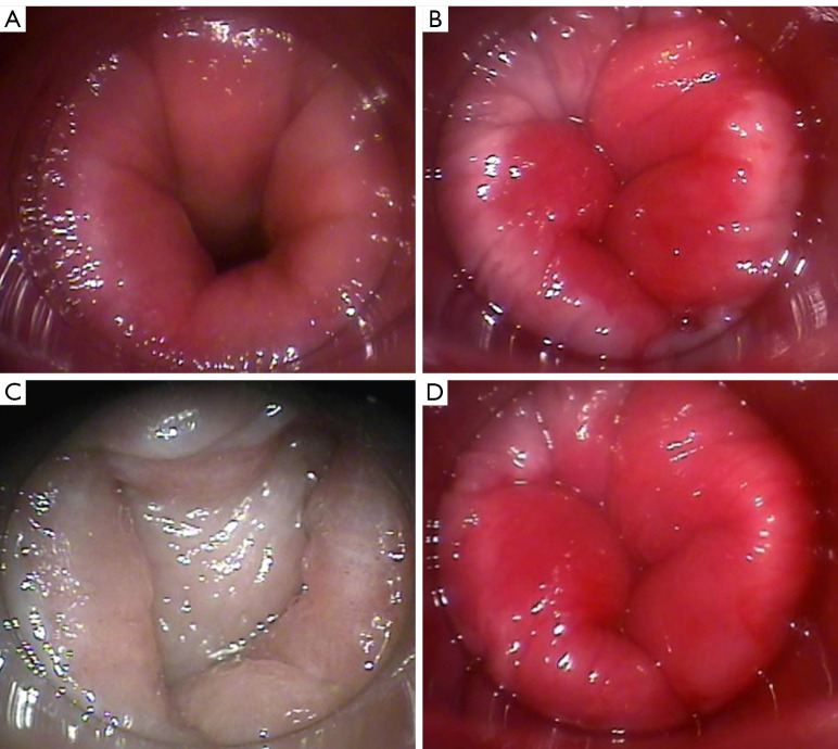
Figure 2.
Images showing the assessment method and morphology of the mucosa at the lower margin of the rectum using the clear plastic proctoscope. (A) Frontal view of rectal lumen when the proctoscope is deeply inserted. (B) Herrmann line when the proctoscope was pulled back to the dentate line. (C,D) Evaluation of the lumen at the end of the rectum: (C) lumen positive; (D) lumen negative.

