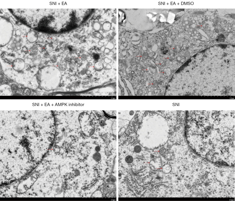
Figure 6.
Autophagosomes were observed using electron microscopy. Rat spinal cord samples were collected from the four groups (SNI + EA, SNI + EA + DMSO, SNI + EA + AMPK inhibitor, and SNI) on day 14 post-surgery following quantification of the paw withdrawal threshold. Electroacupuncture-induced autophagosome formation was observed in the spinal microglial cells, which was inhibited by intrathecal injection of the AMPK inhibitor. Autophagosomes were represented by the red arrows. Scale bar, 2 µm. SNI, spared nerve injury; EA, electroacupuncture; AMPK, AMP-activated protein kinase; DMSO, dimethylsulfoxide.

