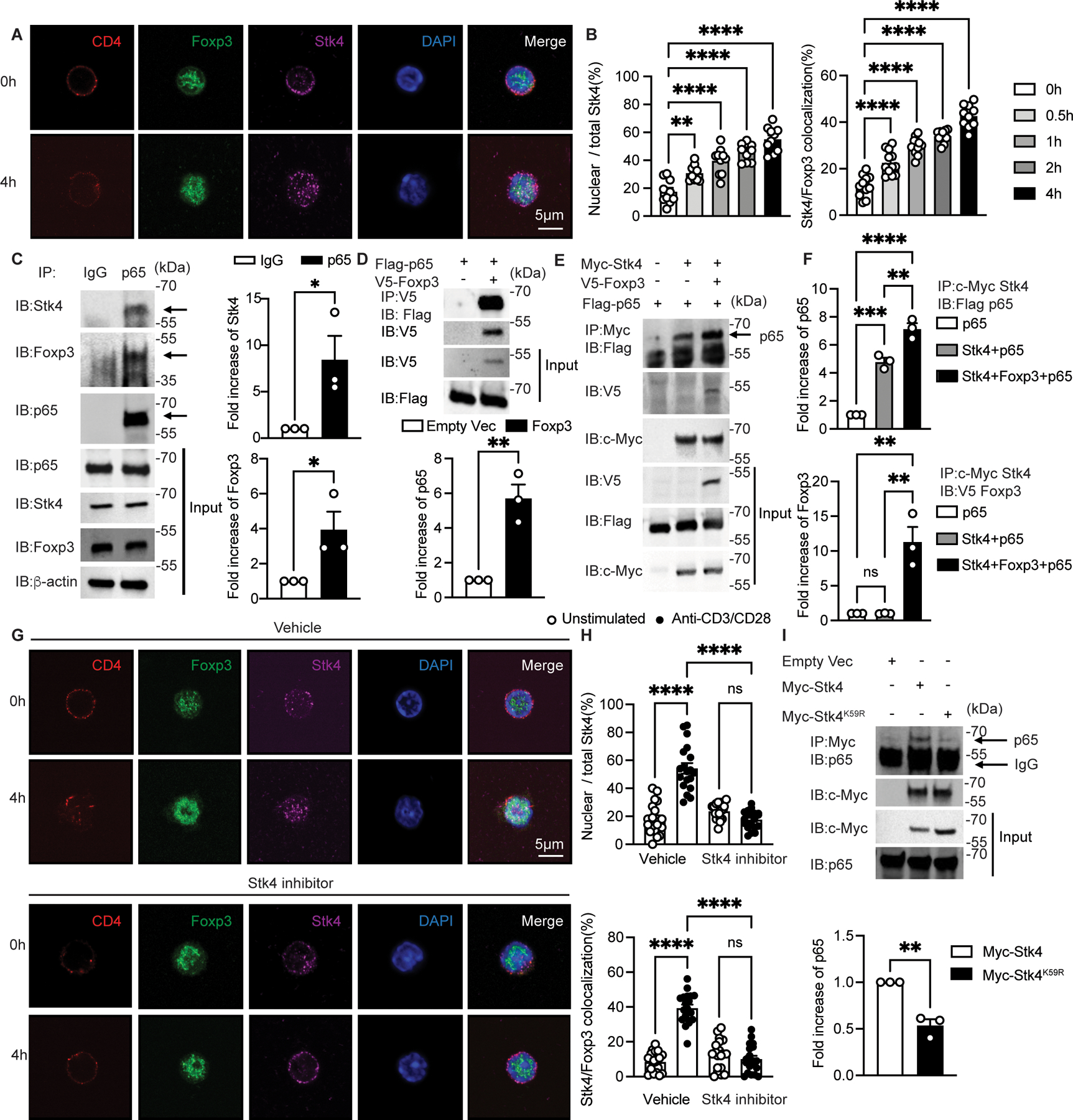
Figure 3. TCR activation stimulates Stk4 nucleus translocation and colocalization with Foxp3.

(A and B) Confocal microscopic analysis of CD4, Foxp3, STK4, DAPI and Merge (A), and ratios of nuclear/total Stk4 and frequencies of Stk4 and Foxp3 co-localization (B) in Foxp3YFPCre Treg cell cultures either unstimulated or stimulated with anti-CD3+anti-CD28 mAbs as indicated (n=10 per group). (C) Immunoblot analysis of Stk4 and Foxp3 association with p65 in Foxp3YFPCre Treg cells that were stimulated overnight with anti-CD3/CD28. Cell lysates were immunoprecipitated and immunoblotted with the indicated antibodies. (D and E) Immunoblot analysis of Stk4 and Foxp3 association with p65 in HEK293T cells transfected with the respective plasmids. Cell lysates were immunoprecipitated with anti-V5 mAb (specific for V5-tagged Foxp3) (D) or anti-c-Myc mAb (E), then immunoblotted with the indicated antibodies. (F) densitometric analysis of immunoblots of p65 (upper panel) and Foxp3 (lower panel) shown in (E). (G and H) Confocal microscopic analysis (G) and frequencies (H) of CD4, Foxp3, STK4, DAPI and Merge, ratios of nuclear/total Stk4 and frequencies of Stk4 and Foxp3 co-localization in Foxp3YFPCre Treg cells either unstimulated or stimulated with anti-CD3+anti-CD28 mAbs without or with the Stk4 kinase inhibitor XMU-MP-1(n=19 per group). (I) Stk4-p65 association is Stk4 kinase activity-dependent. Immunoblot analysis of Stk4-p65 association in HEK293T cells transfected with plasmids encoding either Stk4 kinase-competent (Stk4) or deficient (Stk4K59R) proteins. Each point represents one cell for confocal studies and one blot for immunoblot analyses. Error bars indicate the standard error of the means (s.e.m). Statistical tests: one-way ANOVA with post-test analysis (B and F), two-way ANOVA with post- test analysis (H) and Student’s unpaired two tailed t test (C,D,I). *, P<0.05,**, P<0.01, ***, P<0.005, ****, P<0.0001.
