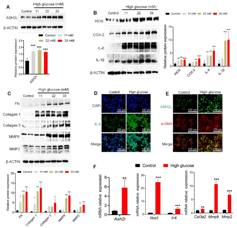
Figure 1.
High glucose activates ASH2L aligned with fibrosis and inflammation in mesangial cells. (A–C) Western blot analysis of ASH2L, fibrosis markers (FN, Collagen 1, Collagen 3, MMP9, and MMP2), and inflammatory mediators (iNOS, COX-2, IL-6, and IL-1β) expression in SV40-MES-13 cells treated with 11 mM, 22 mM, and 33 mM D-glucose for 24 h. Data from at least three independent experiments are shown as mean ± S.D, * p < 0.05, ** p < 0.01, and *** p < 0.001 compared with the control group. (D,E) Immunofluorescence staining of IL-6, ASH2L, and α-SMA in SV40-MES-13 cells treated with or without 33 mM D-glucose (high glucose) for 24 h. (F) Quantitative RT-PCR analyses of the relative mRNA levels of Ash2l, fibrosis-associated markers (Col3a2, Mmp9, and Mmp2), and inflammatory mediators (Nos2 and Il-6) in SV40-MES-13 cells treated with or without 33 mM D-glucose. Data from at least three independent experiments are shown as mean ± S.D, ** p < 0.01, and *** p < 0.001 compared with the control group.

