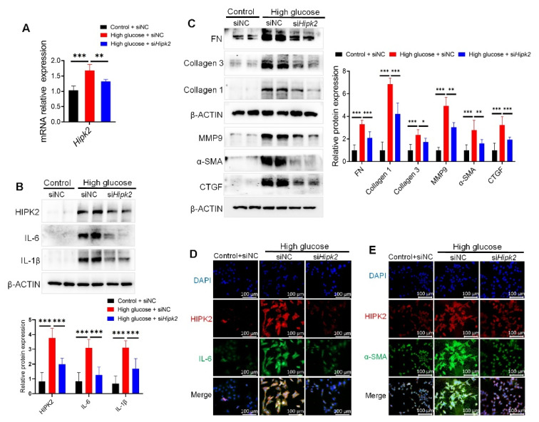
Figure 5.
Loss of HIPK2 decreases fibrosis and inflammation in mesangial cells. (A) Quantitative RT-PCR analyses of Hipk2 in SV40-MES-13 cells treated with or without 33 mM high glucose for 24 h after transfection with negative control (siNC) or HIPK2 small interfering RNA (siHipk2). Data from at least three independent experiments are shown as mean ± S.D, ** p < 0.01, and *** p < 0.001. (B,C) Western blot analysis of HIPK2, inflammatory mediators (IL-6 and IL-1β), and fibrosis markers (FN, Collagen 3, Collagen 1, MMP9, α-SMA, and CTGF) expression in SV40-MES-13 cells treated with or without 33 mM high glucose for 24 h after transfection with siNC or siHipk2. Data from at least three independent experiments are shown as mean ± S.D, * p < 0.05, ** p < 0.01 and *** p < 0.001. (D,E) Immunofluorescence staining of HIPK2, α-SMA, and IL-6 in SV40-MES-13 cells treated with or without 33 mM D-glucose for 24 h after transfection with siNC or siHipk2.

