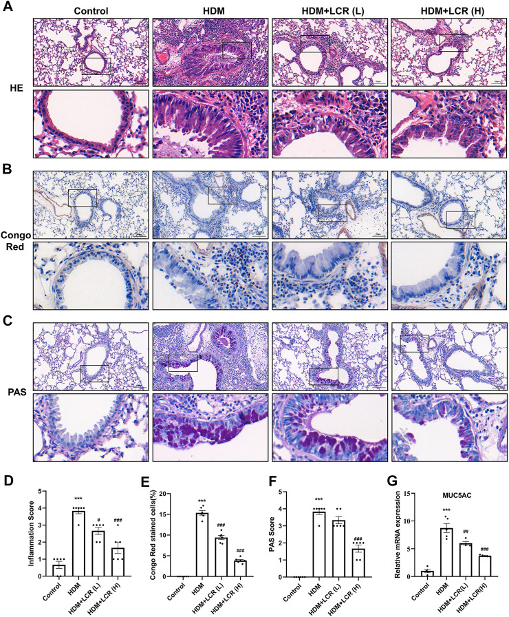
FIGURE 4.
Lonicerin alleviated inflammatory cell infiltration and mucus hypersecretion in the lungs of asthmatic mice. (A) HE staining was performed to evaluate airway inflammatory infiltration. (B) Lung eosinophils detected by Congo red staining. (C) PAS staining was performed to show mucus production (scale bar, 100 µm). (D–F) The corresponding quantification histograms of (A–C), respectively. (n = 6). (G) Detection of MUC5AC mRNA expression in lung tissues of different groups of mice by qPCR. (n = 4–5). Data are expressed as mean ± SEM. Differences between groups were determined with one‐way ANOVA followed by the Tukey’s post hoc test. *p < 0.05, **p < 0.01, ***p < 0.001 vs. Control group; #p < 0.05, ##p < 0.01, ###p < 0.001 vs. HDM group.

