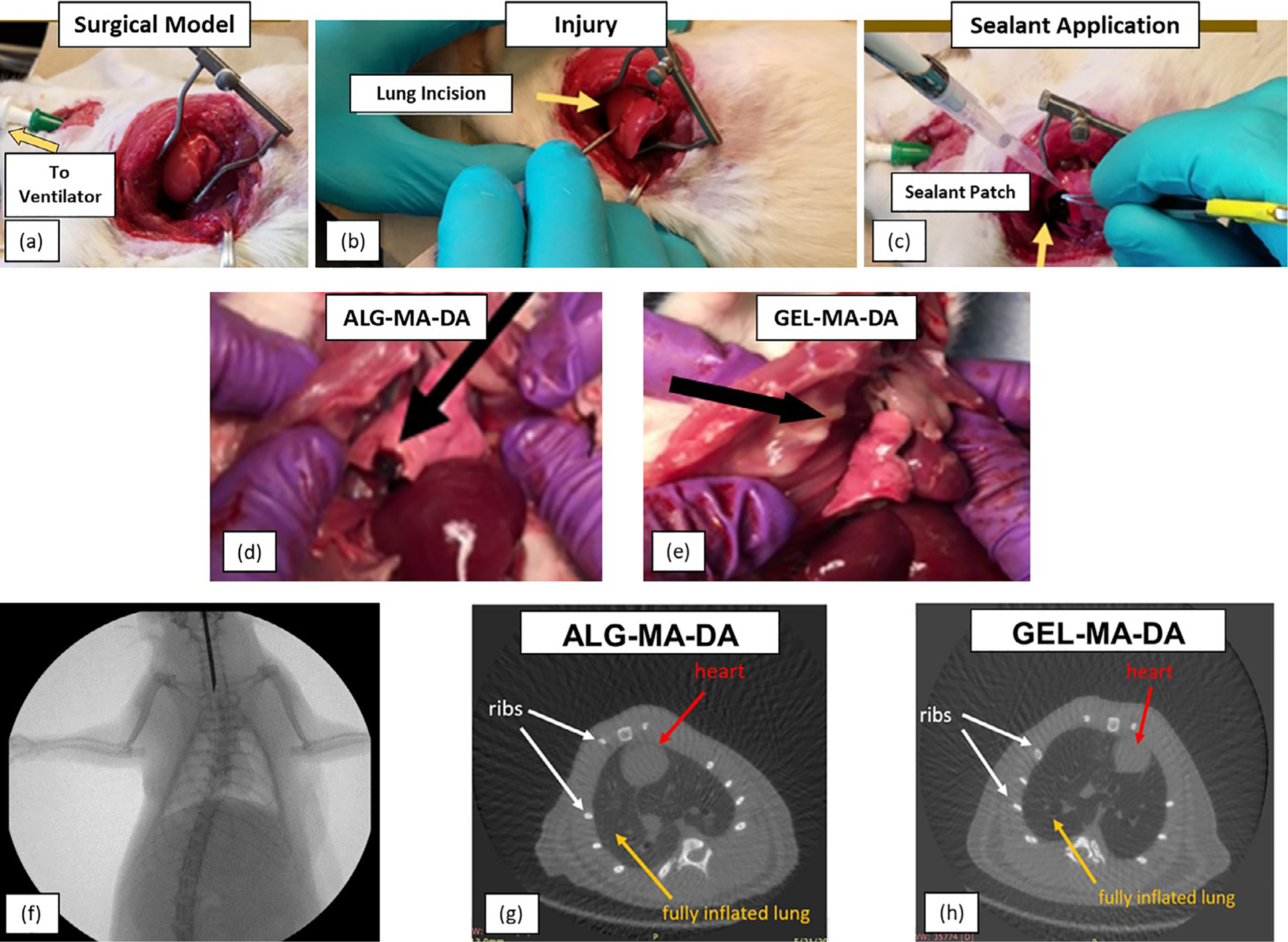
Fig. 7. In vivo model for testing pleural sealant.

(a) Anesthetized, intubated and mechanically ventilated rats underwent thoracotomy to expose the right lung. (b) Injury is induced by puncture with an 18 g needle and leak of air bubbles observed to confirm injury. (c) The sealant is applied with cessation of air leak. (d) Necropsy of rat 1 week or month post operatively. Residual ALG-MA-DA patch material was observed in rats receiving ALG-MA-DA patch (black arrow). No GEL-MA-DA material was grossly visible in rats receiving GEL-MA-DA in situ formed hydrogel application. In neither case was obvious inflammatory reaction or evidence of sealant observed on the parietal pleura/chest wall. High power images are included for panels a-e. (f) Representative fluoroscopy after 1 week demonstrates clear lungs and no obvious pneumothorax. (g) and (h) Representative CT scans at 1 month demonstrates clear lungs and no pneumothorax. White arrows point the ribs; yellow arrows demonstrate a fully inflated lung and red arrows show the position of the heart.
