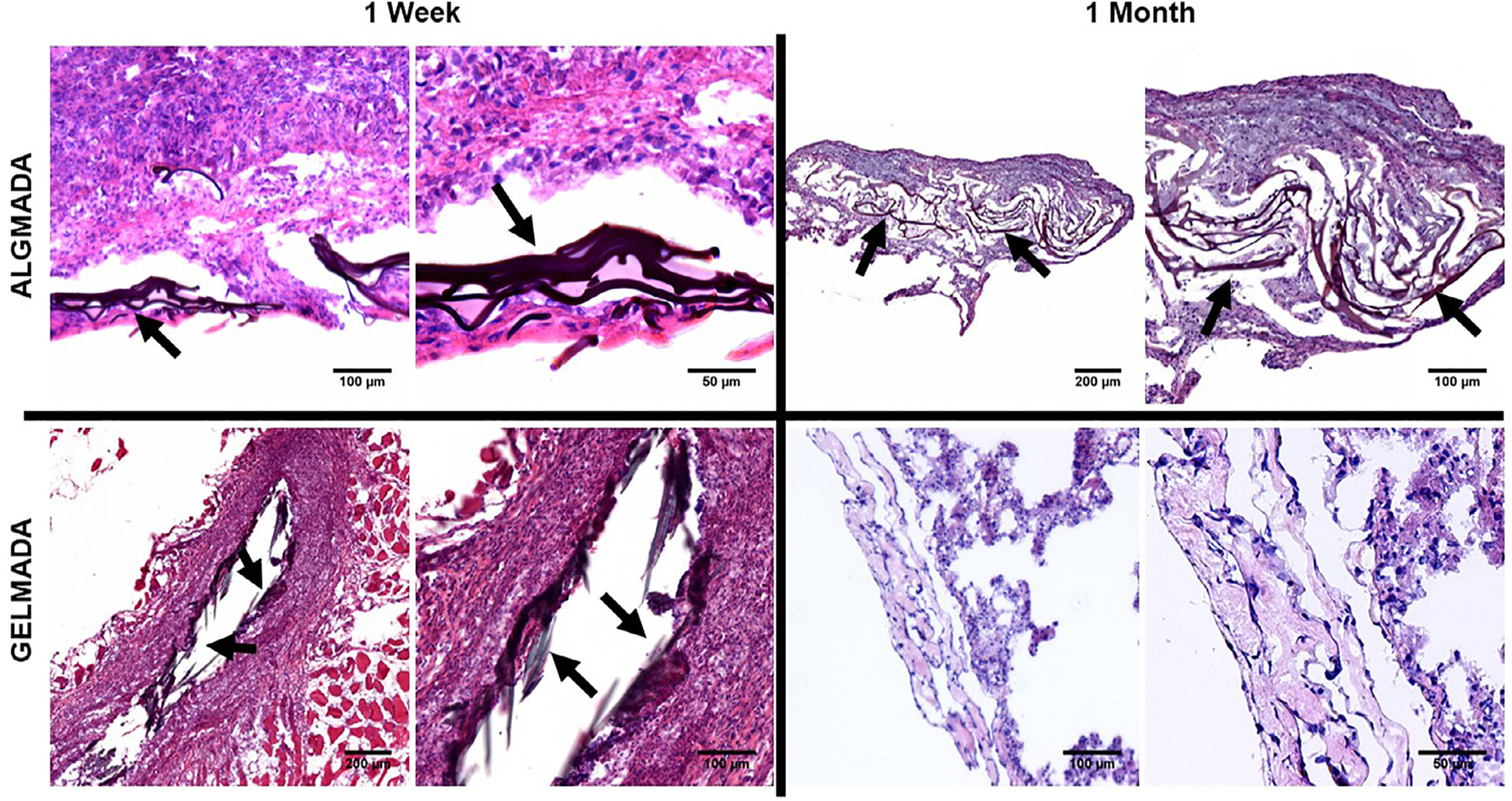
Fig. 8.

H and E stained sections demonstrate good wound repair, no obvious histologic inflammation, and residual sealant (arrows) for 1 week GEL-MA-DA and both 1 week and 1 month ALG-MA-DA. The one week GEL-MA-DA image depicts residual sealant in what appears to be the needle injury tract. Inset depict higher power magnifications of the respective designated areas.
