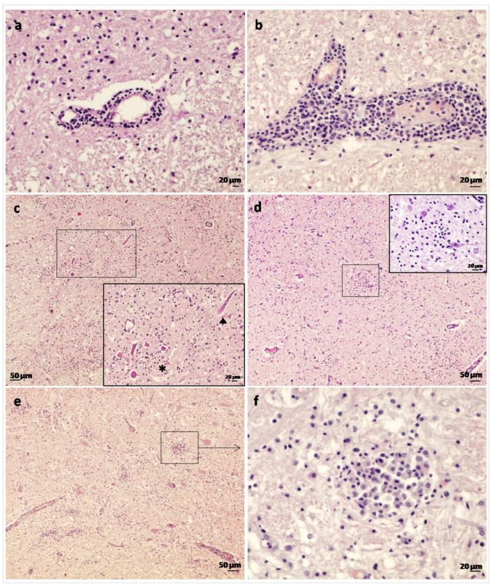
Figure 1.
Histological lesions in goats affected by the Spanish goat encephalitis virus (SGEV) as shown by HE staining. (a) Spinal cord: thin perivascular cuff comprising only one to two layers of small mononuclear inflammatory cells. (b) Midbrain: thick perivascular cuff which consisted of lymphocytes, plasma cells and macrophages. (c) Spinal cord: widespread gliosis in grey matter. Inset: Area containing several necrotic neurons (*) and axonal tumefaction (arrowhead). (d) Midbrain: occasional glial focus present. Inset: Detail of glial focus composed of microglial cells. (e) Cerebellar peduncles: severe widespread inflammation and multiple glial foci in the vaccinated goat (No. 3). (f) Cerebellar peduncles: detail of a glial focus comprised primarily of macrophages with fewer numbers of lymphocytes and glial cells.

