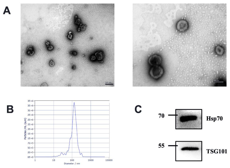
Figure 1.
Characterization of EVs from bovine milk (A) Transmission electron microscopy observation of milk-derived EVs, left, scale bar size 200 nm; right, scale bar size 100 nm; (B) Nanoparticle tracking analysis showing particle size distributions of milk-derived EVs. (C) Western blotting of exosome common markers TSG101 and Hsp70 in milk-derived EVs (original western blot figures in Figure S1).

