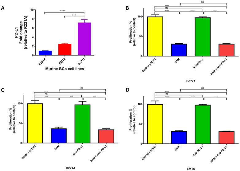
Figure 2.
PD-L1 expression and effect of PD-L1 intracellular signaling on cell proliferation of murine BCa cells. (A) Expression of PD-L1 in murine BCa cell lines analyzed by RT-qPCR. The fold change was relative to the expression of R221A. (B–D) Effect of SAM and anti-PD-L1 antibody on proliferation of murine BCa cells. (B) Eo771 (4 × 10⁴), (C) R221A (1 × 10⁴), and (D) EMT6 (4 × 10⁴) cells were seeded in 6-well plates and were added to rPD-1 (0.2 μg/mL, day 3). The cells were treated with either control (only rPD-1), SAM (200 μM, day 2, 3, 4), anti-PD-L1 antibody (50 μg/mL, day 4), or SAM and anti-PD-L1 in combination. The results are the mean of at least three independent experiments. Proliferation is represented as the percentage proportional to the control (± SEM). Statistical significance was determined by one-way ANOVA in GraphPad prism. Significance values are represented by asterisks (ns; not significant; *** p < 0.001; **** p < 0.0001).

