Figure 2.
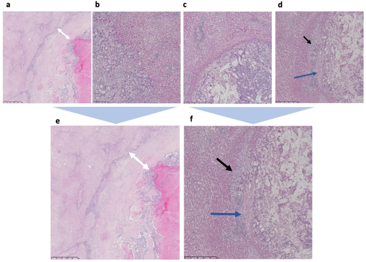
Histopathological growth patterns in a four-tier and two-tier system (H&E images, original data). (a) Low magnification of desmoplastic HGP, with white arrows marking the thick fibrous rim that separates the tumor from hepatocytes. (b) Low magnification of tumor–liver interface in replacement HGP with cancer cells in continuity with normal hepatocytes. Cancer cells form solid nests and trabeculae and there is no glandular differentiation. (c) Low magnification of pushing HGP with sharp interface between tumor cells and adjacent hepatocytes, without desmoplastic rim or tumor cells invading into liver tissue. (d) Low magnification of mixed/heterogeneous growth with replacement growth pattern (black arrow) and desmoplastic rim (blue arrow). (e) higher resolution of desmoplastic HGP, with white arrows marking the thick fibrous rim that separates the tumor from hepatocytes. (f) higher resolution of of mixed/heterogeneous growth with replacement growth pattern (black arrow) and desmoplastic rim (blue arrow). Images are provided by the authors’ clinical series.

