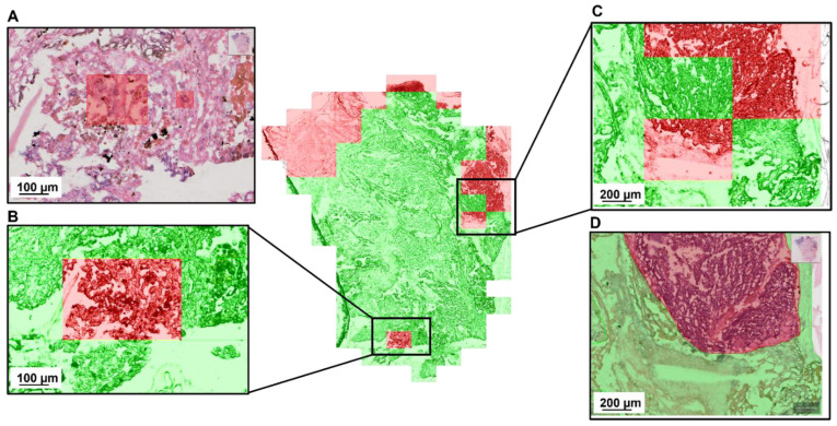
Figure 4.
Detailed evaluation of the 3d CNN-based tumor classification with a false positive (B) and a false negative (C) sample. Both regions were given to a pathologist for reevaluation. The annotation (D) was confirmed by the second pathologist. The red regions in (A) mark features that the second pathologist labelled as ambiguous.

