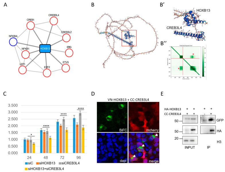
Figure 6.
CREB3L4 is a novel cofactor of HOXB13 that can promote the proliferation of prostate-cancer-derived PC-3 cells. (A) Interactome of the HOXB13 enriched cluster involved in prostate cancer. Candidate cofactors have a known tumor suppression gene (TSG (blue circle)) or oncogenic (red circle) function. (B) AlphaFold prediction of the interaction between CREB3L4 and HOXB13. (B’) Enlargement of the interaction interfaces between the homeodomain (HD) of HOXB13 and the alpha helix of CREB3L4. (B”) Prediction score of intra-domain interactions of HOXB13 (upper dark green box; corresponds to the HD) and CREB3L4 (lower dark green box; corresponds to the alpha helix) and extra-domain interactions between the HD of HOXB13 and the alpha helix of CREB3L4 (green boxes highlighted by a red star). The first 0-280 residues correspond to HOXB13, and the following 280-700 residues correspond to CREB3L4. (C) xCELLigence assay of PC-3 cells transfected with the different siRNAs as indicated. siC = siRNA control (see also Materials and Methods). Measures were performed at different time points post-transfection and resulted from three independent biological replicates. Two-way ANOVA with Tukey’s multiple comparisons; * p < 0.05 and **** p < 0.0001. (D) Illustrative confocal picture of BiFC (green) of VN-HOXB13 and CC-CREB3L4 in fixed HEK293T cells. The mCherry reporter (red) stains show transfection and DAPI (blue) indicates cell nuclei. A typical enrichment was observed in dividing nuclei (white arrows). (E) Co-IP of HA-tagged HOXB13 and CC-CREB3L4 co-expressed in HEK293T cells. The Co-IP was performed with anti-HA, and CREB3L4 was revealed with anti-GFP, which recognizes the CC fragment (see Materials and Methods). “+” and “−”, respectively, denote the co-transfection or lack of co-transfection of HA-HOXB13 with CC-CREB3L4. The protein size scale is indicated on the left side (KDa). Confocal and Western blot pictures are illustrative of two independent biological replicates.

