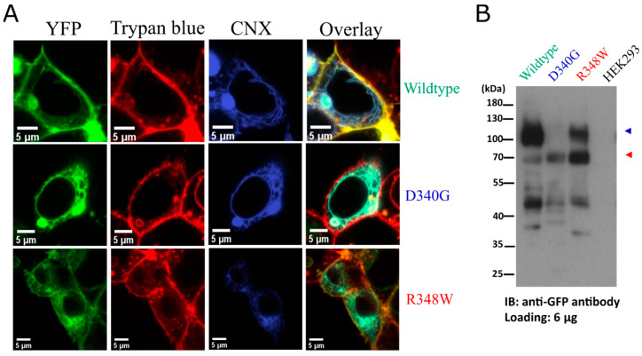
Figure 2.
(A) Representative images of YFP-tagged wildtype OCT3 (top row), OCT3-D340G (middle row) and OCT3-R348W (bottom row) were captured by confocal microscopy to visualize the YFP-tagged transporter (left hand column), trypan blue (second column) and mTagBFP-tagged calnexin (CNX; third column). The overlay is shown in the right-hand column. (B) Western blot of lysates (6 µg) prepared from HEK293 cells expressing YFP-tagged wildtype OCT3, OCT3-D340G, OCT-R348W and from untransfected HEK293 cells. The red and the blue arrow indicate the position of the ER-resident core-glycosylated transporter and mature glycosylated transporter, respectively.

