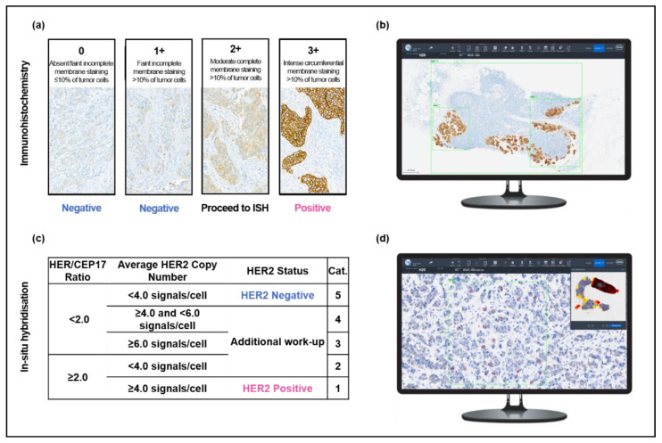
Figure 1.
Overview of the study protocol for HER2 evaluation. Pathologists performed manual, light microscopic evaluation of HER2 (a) immunohistochemistry and (c) in situ hybridization in accordance with the 2018 ASCO/CAP guidelines. Slides were also analyzed with the support of two AI algorithms: AI-assisted immunohistochemistry analysis (b) was performed by placing three regions of interest (green) over tumor tissue and (d) AI-assisted in situ hybridization was analyzed by selecting a region of interest (green) within an area of high HER2 expression, as indicated by the heatmap (upper right). Within the region of interest, the algorithm calculated the HER2 and CEP17 signals for 20 cells (red).

