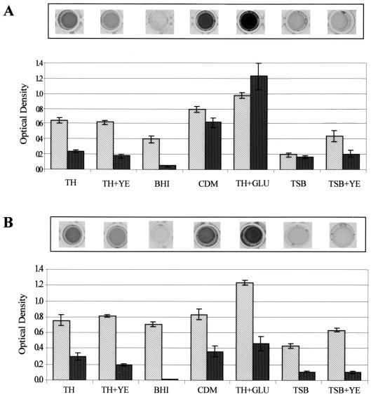
FIG. 1.
Bacterial growth and biofilm formation of S. parasanguis FW213 under different growth conditions. Cells grown in TH to early stationary growth phase were washed once in sterile water and subcultured (1:200) into either TH, TH+YE, BHI broth, CDM, TH+Glu, TSB, or TSB+YE. These cultures were then grown in polystyrene microtiter dishes at 37°C for 16 h either aerobically in 5% CO2 (A) or anaerobically (B). Growth (light bars) and biofilm formation (dark bars) were quantified as optical density at 490 and 562 nm, respectively. Assays were performed in quadruplicate; mean values and standard deviations are shown. A representative row of crystal violet-stained microtiter plate wells is shown above each graph.

