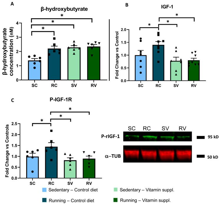
Figure 3.
Vitamin supplementation blocks IGF1 increase in running animals. (A) Colorimetric assay for serum β-hydroxybutyrate. Running induced an increase in circulating β-hydroxybutyrate with respect to the sedentary condition (RC, n = 6; 2.20 ± 0.17 nM; vs. SC, n = 6; 1.37 ± 0.13 nM; two-way ANOVA, F = 7.17, Holm–Sidak method, p < 0.001). Vitamin supplementation caused an increase of β-hydroxybutyrate levels in sedentary animals (SV, n = 6; 2.29 ± 0.12 nM; p < 0.001), but no further increase was observed after free running. (B) ELISA for serum IGF-1. Serum IGF-1 levels were markedly increased in exercised rats as compared to sedentary animals fed with the control diet (n = 8 and n = 7, respectively; two-way ANOVA, F = 1.92, Holm–Sidak method, p = 0.047). Vitamin supplementation completely abolished the IGF-1 increase induced by physical exercise, without any effect in sedentary animals (n = 8 and n = 7, p = 0.003 and p = 0.006, respectively). (C) Western blot for pIGF-1 receptor in the visual cortex. Physical activity led to a marked increase of phosphorylated rIG1-1 in the visual cortex of RC (n = 6) vs. SC (n = 6) animals; vitamin supplementation completely blocked this increase, with RV rats (n = 6) displaying levels of phosphorylated IGF-1 significantly lower than those displayed by RC rats (two-way ANOVA, F = 1.74, Holm–Sidak method, p = 0.037, p = 0.013, respectively). No effect was found in the visual cortex of sedentary animals fed with the diet supplemented with antioxidants (n = 6) (SC vs. SV, p = 0.407). The right panel shows representative bands for all groups. To calculate the fold change for both (B,C) panels, each individual value was normalized, dividing it by the mean value of the SC control group. * indicates statistically significant differences; error bars represent SEM.

