Figure 5.
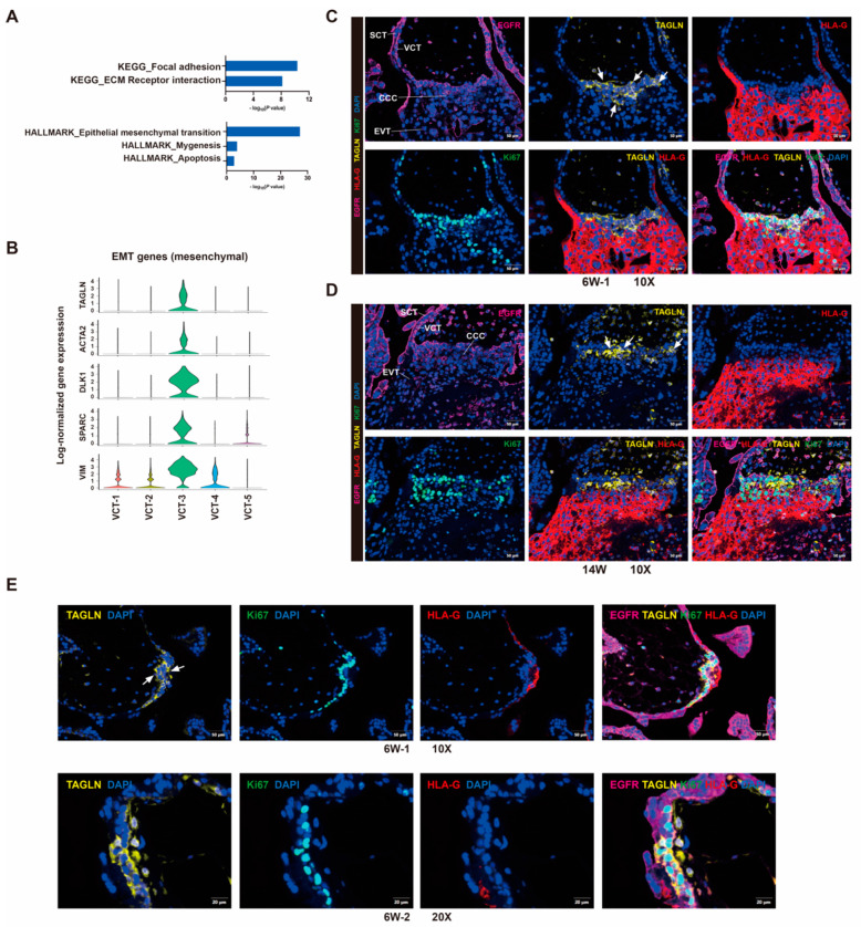
Human trophoblast progenitor cells contribute to EVT lineages. (A) Selected top categories from KEGG and Cancer Hallmark analysis of differentially expressed genes in VCT-3. (B) Violin plots showing the relative expression of EMT genes in each VCT cluster. (C,D) Immunofluorescence staining for the indicated VCT-3 marker TAGLN, trophoblast marker EGFR, EVT marker HLA-G, proliferative cell marker Ki67 in cytotrophoblast cell columns structures of 6 weeks of gestation (C) and 14 weeks of gestation (D) placental tissue. Scale bars, 50 μm (E) Immunofluorescence staining for TAGLN in the two-layer cell structure of 6 weeks of gestation placental villi. The white arrowheads indicated VCT-3 cells, which were TAGLN and EGFR double positive, and HLA-G negative. Scale bars, 50 μm for 10× and 20 μm for 20×.

