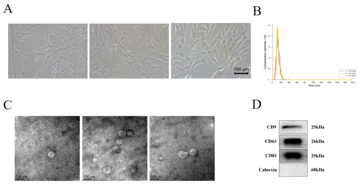
Figure 1.
Identification of human umbilical cord MSCs (hUC-MSCs) and MSC-derived extracellular vesicles (MSC-EVs). (A) Light microscope image of hUC-MSCs (20×). (B) Diameters and concentration of MSC-EVs were detected by nanoparticle trafficking analysis. (C) Identification of MSC-EVs by transmission electron micrograph. (D) Western blot showed the protein level of MSC-EVs CD9, CD63, CD81, and Calnexin.

