Figure 6.
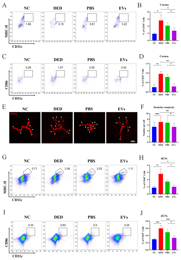
Phenotype change of DCs in DED mice was suppressed by topical application of MSC-EVs. To evaluate the effects of MSC-EVs on the phenotype of DCs, we detected the immunophenotype and morphological characters of DCs. Representative flow cytometry plots showing the frequency of corneal CD11c+ MHC-II+ DCs (A) and CD11C+ CD86+ DCs (C) in mice corneas after treatment with topical MSC-EVs or PBS. Bar charts showing the frequency of corneal CD11c+ MHC-II+ DCs (B) and CD11C+ CD86+ DCs (D) in mice corneas after treatment with topical MSC-EVs or PBS. (E) Representative micrographs (20×) of the dendritic complexity (white arrowhead) of DCs in the central and peripheral cornea of each treatment group. (F) Bar charts showing the dendritic complexity in the central and peripheral cornea after treatment with topical MSC-EVs or PBS. Representative flow cytometry plots showing the frequency of CD11c+ MHC-II+ DCs (G) and CD11C+ CD86+ DCs (I) in the dLNs of MSC-EVs or PBS treated DED mice on day 14. Bar charts showing the frequency of CD11c+ MHC-II+ DCs (H) and CD11C+ CD86+ DCs (J) in the dLNs of each treatment group. Three independent experiments (n = 8 mice per group) were pooled. Data are presented as mean ± SD of three independent experiments, each consisting of eight mice per group (ns: not significant, * p < 0.05, ** p < 0.01, *** p < 0.001).

