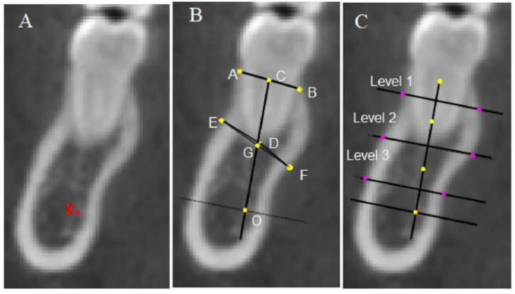
Figure 6.
Landmarks and linear measurements of a mandibular posterior tooth. (A) X: Top point of the mandibular alveolar neural tube. (B) A, B: Buccal alveolar crest and lingual alveolar crest; C: midpoint of the AB line. D: Root apex point. E: The point on the buccal wall closest to the root apex point; F: the point on the lingual wall closest to the root apex point; G: midpoint of EF line; O: the foot point of a line perpendicular to the CG line at point X; the CO line represents the long axis of the alveolar bone. The length of the CO line is the alveolar height. (C) The long axis of the alveolar bone (CO line) is divided into thirds (shown by the yellow dots), which mark the midpoint of each third line as purple dots. Three lines at different levels are drawn perpendicular to the long axis of the alveolar bone and extend from the buccal to the lingual cortical plate. The distance between the two plates represents the alveolar thickness at level 1 (coronal third), level 2 (middle third), and level 3 (apical third), respectively.

