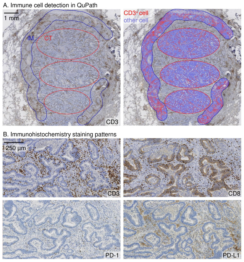
Figure 1.
Immune cell density analysis and immunohistochemistry staining patterns in a pulmonary metastasis of colorectal cancer. (A). Analysis of immune cell density in the representative sites of the tumour centre (CT) and the invasive margin (IM) sites. The width of the invasive margin was 720 µm spanning 360 µm into the tumour and 360 μm into the healthy tissue. The immune cell density analyses for CD3, CD8 and PD-1 were done in QuPath bioimage software. PD-L1 expression was scored manually. (B). Examples of CD3, CD8, PD-1, and PD-L1 staining patterns are represented in the respective site of the tumour.

