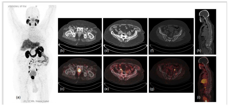
Figure 3.
87-year-old male diagnosed with adenocarcinoma of prostate (Gleason score 7) in 2017 was heavily pre-treated: ADT with Pamorelin + Bicalutamide followed by Abiraterone and Enzalutamide. Nadir PSA was 0.016 ng/mL during the treatment and biochemical progression was observed for 6 months following. He presented to the doctor’s office with shortness of breath, limp legs, weakness and leg pain, his serum PSA was 94.7 ng/mL, and he was referred for PSMA PET CT scan. 68Ga-PSMA-11 PET CT scan showed prostatomegaly invading the urinary bladder wall with PSMA expressing irregular enhancing SOL involving almost entire gland. PSMA expressing multiple metastatic pelvic and retroperitoneal lymph nodes and PSMA expressing multiple sclerotic metastatic skeletal lesions showed disease progression as compared to previous PSMA PET-CT scan of 2019. (a)-MIP image, (b–i)-CT and fused PET-CT trans-axial and sagittal images.

