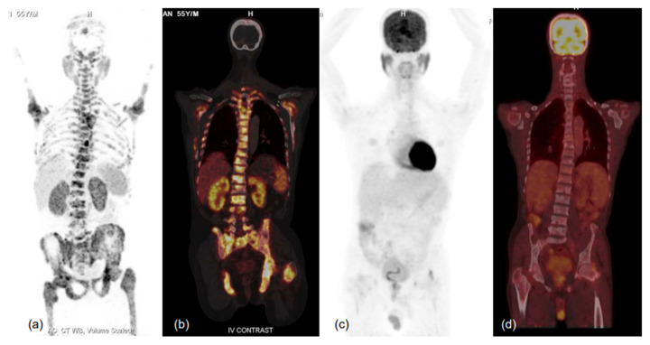
Figure 6.
MIP images of 68Ga-PSMA-11 (a,b) and FDG (c,d) PET-CT scans and corresponding fused coronal images (b-68Ga-PSMA-11 and d-FDG PET-CT scans) in bone window showing PSMA-expressing multiple sclerotic and marrow lesions involving almost the entire axial and proximal appendicular skeleton, while no significant FDG uptake (metabolic activity) is evident in the lesions. Such patients respond well to therapy, especially PSMA-targeting radionuclide therapy, and demonstrate relatively favourable prognoses in terms of survival and quality of life benefits. Thus, dual-tracer PET using FDG and 68Ga-PSMA-11 helps in patient selection for PSMA peptide receptor radionuclide therapy (PRLT) and in predicting treatment outcomes. He was scheduled for 177Lu-PSMA-617 therapy, but unfortunately could not attend due to logistical constraints.

