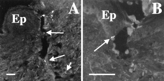
FIG. 1.
Deposition of H. ducreyi in skin. Sections from tissue biopsied immediately postinoculation were stained with polyclonal anti-H. ducreyi antiserum and MAb BB11. (A) Puncture wound made by inoculation device. Arrows indicate bacteria along puncture wound. (B) Bacteria (arrow) at the epidermis with keratinocytes. Ep, epidermis. Bars, 50 μm.

