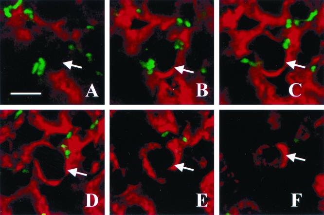
FIG. 4.
Optical sectioning through a pustule. Panels A through F represent, in order, images taken in 1-μm steps through a section stained with polyclonal anti-H. ducreyi antiserum (green) and anti-PMN elastase MAb (red). The arrow in each panel points to one edge of the PMN. Note the bacteria on the outside but not within the PMN. Bar, 5 μm.

