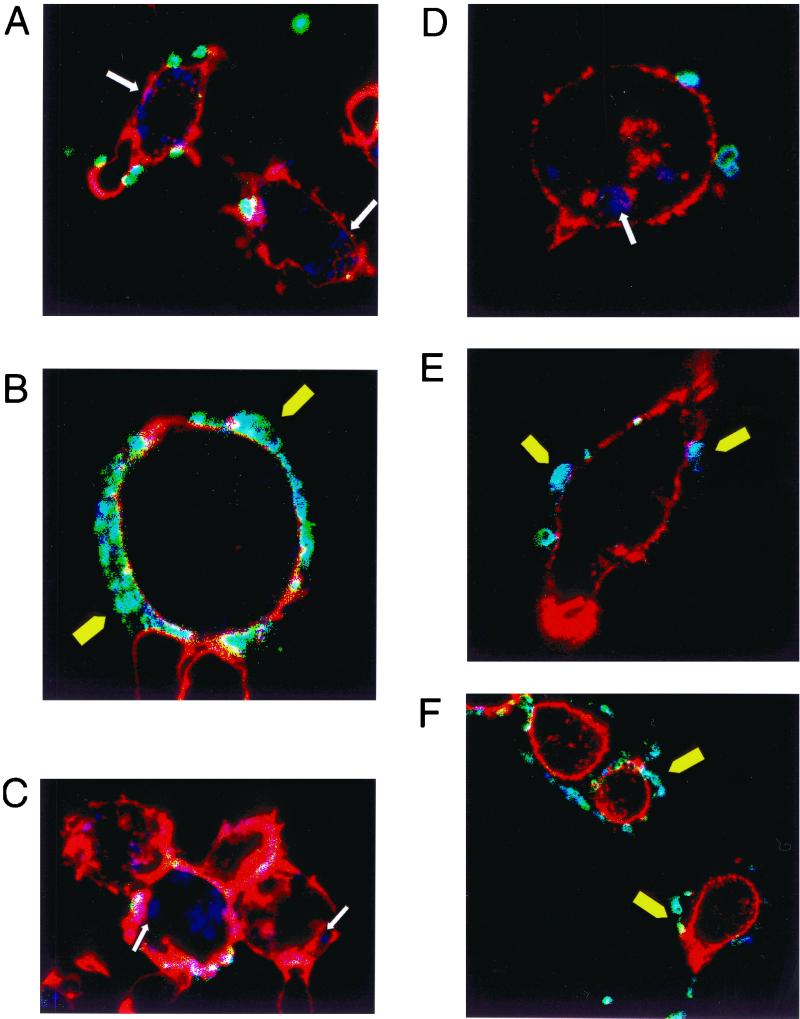
FIG. 2.
(A) Confocal picture of 5-day-derived macrophages infected with N303. (B) Confocal picture of 4-day-derived macrophages infected with P12. (C) Confocal picture of 2-day-derived macrophages infected with PAI. (D) Confocal picture of a J774a cell infected with N303. (E and F) Confocal pictures of J774a cells infected with P12 followed by an infection with N303; only the gonococci are stained. White arrow shows intracellular bacteria; yellow arrowheads show extracellular bacteria.

