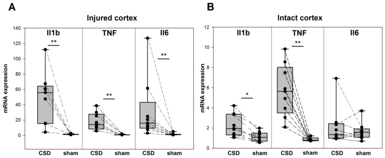
Figure 3.
Proinflammatory cytokine expression following a single CSD in the injured and intact regions of the cerebral cortex. Levels of Il1b, TNF, and Il6 mRNA (medians with interquartile ranges and individual data from each rat, n = 9) in the perilesional tissue of the somatosensory cortex (A) and in the intact tissue of the frontal cortex (B) of the lesioned (CSD) and opposite (sham) hemispheres. * p < 0.017 and ** p < 0.0085—ipsilesional versus sham-treated cortical regions (post hoc Wilcoxon test with Bonferroni correction).

