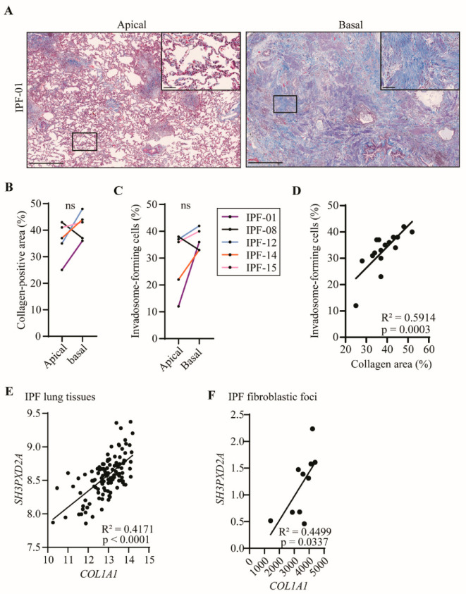
Figure 3.
Invadosomes are found where collagen is heavily produced. (A) Images of apical and basal lung tissue sections stained for collagen with Masson’s trichrome from the same IPF patient showing different levels of fibrosis maturity. Scale bar = 1000 µm or 100 µm (zoom). For five patients, apical and basal lung samples were collected and (B) percentage of collagen-positive area as well as (C) percentage of fibroblast-forming invadosomes were determined. (D) Percentage of collagen-positive areas in lung tissues versus percentage of invadosome formation (n = 17). TKS5 (SH3PXD2A) relative gene expression was correlated with COL1A1 expression in (E) IPF lung tissue (n = 119) and in (F) IPF fibroblastic foci (n = 10) using GSE32537 and GSE169500 cohorts, respectively.

