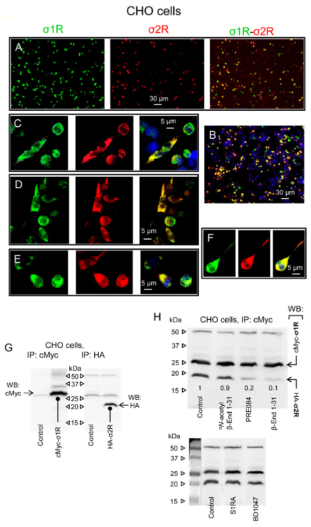
Figure 6.

Physical interactions between σ1Rs and σ2Rs. Effect of β-End 1–31 and αN-acetyl derivative. (A) Single-wavelength and merged images of CHO cells cotransfected with cMyc-σ1R and HA-σ2R. The cells were fixed 48 h after transfection and analyzed by confocal laser scanning microscopy. Transient expression of proteins within the transfected cells was detected with anti-cMyc (green) and anti-HA (red) antibodies (original magnification 10×). The fluorescent signals reflect the expression of the proteins of interest. Scale bar: 30 µm. (B) Nuclei stained with DAPI (blue). (C–F) Single-wavelength and merged images are enlargements of individual cells. Scale bar: 5 µm. Lower panels: (G) Immunoprecipitation (IP) of tagged sigma receptor types from cotransfected CHO cells. Biotin-conjugated anti-cMyc and anti-HA antibodies precipitated sigma receptors as detected by Western blot analysis using antibodies that recognize either tag of the sigma receptor proteins. (H) σ1R/σ2R coprecipitates from cultures incubated with β-End 1–31 (1 nM), αN-acetyl β-End 1–31 (1 nM), the σ1R agonist PRE084 (100 nM) and the σ1R antagonists S1RA (100 nM) and BD1047 (100 nM). The control was assigned an arbitrary value of 1, and the changes observed in the test groups were referred to as the control. Representative blots are shown.
