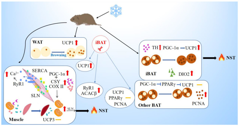
Figure 6.
Schematic model of nonshivering thermogenesis induced by cold and iBAT removal in Brandt’s voles. The box represents cold-induced thermogenesis of tissues, and the circle represents the effect of iBAT removal. Yellow dashes represent no significant change in indicators. Cold acclimation induces browning of WAT, and results in up-regulation in the levels of TH, PGC-1α, UCP1 and DIO2 for thermogenesis in iBAT, but does not affect BAT in other sites except for increased PCNA level. Upregulation of PGC1a would promote mitochondrial biogenesis in the muscle (assisted by COXII & CSY) that will support SLN-mediated thermogenesis. Upregulated RyR1 will increase leak and cytosolic Ca2+ that will activate muscle NST. Cold enhanced thermogenesis indicated by increased expression of SERCA, but with no change in UCP3. IBAT removal induced supplement in WAT browning and muscle metabolism and thermogenesis indicated by increased expression of ACACβ and RYR1. WAT, white adipose tissue; iBAT, interscapular brown adipose tissue; UCP1, uncoupling protein 1; TH, tyrosine hydroxylase; PGC-1α, peroxisome proliferator-activated receptor gamma coactivator 1-alpha; PPARγ, peroxisome proliferator-activated receptor gamma; DIO2, deiodinase iodothyronine type II; PCNA, proliferating cell nuclear antigen; CSY, citrate synthetase; ACACβ, acetyl-CoA carboxylase beta; COX II, cytochrome C oxidase subunit II; SERCA, sarcoplasmic endoplasmic reticulum Ca2+-dependent adenosine triphosphatase; SLN, sarcolipin; RyR1, the type 1 ryanodine receptor; UCP3, uncoupling protein 3; NST, nonshivering thermogenesis.

