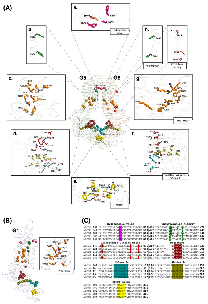
Figure 1.
Motifs and elements in ABCG1 and ABCG5/G8. (A) Stick illustration of structural motifs shown on the crystal structure of ABCG5/G8 (PDB ID: 8CUB). (a–i) In the extracellular region, hydrophobic valve residues are shown in pink; in the TMDs, phenylalanine highway, cholesterol-sensing motif, and polar relay are represented in green, red, and orange, respectively; in the NBDs, signature, Walker A, Walker B, and NPXDF motifs are illustrated in brick red, deep teal, deep olive, and yellow, respectively. * The tyrosin residue labelled in the polar relay and phenylalanine highway is common in these two motifs. (B) The same conserved motifs with comparable colors in the structure of an ABCG1 half transporter. The zoomed-in picture shows the involved residues appear to form a polar relay network on the TMD of an ABCG1 monomer. These residues are highly conserved in mammals (see Supplementary Materials). (C) The sequence alignment illustrates conserved residues of structural motifs shown in panels A and B in all ABCG subfamily members (colors are picked in accordance with the color code in panels (A,B)).

