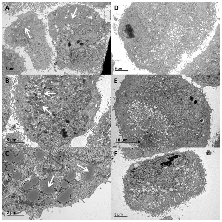
Figure 5.
Lipid droplet resembling structures in hPod exposed to active disease FSGS plasmas. hPod differentiated in T25 culture flasks (Corning) were exposed to FSGS plasmas. Electron microscopy images of hPod exposed to (A) active disease plasma nat1A, (B) post-transplant recurrence plasma rec1B, (C) nat1A plasma zoomed in, (D) remission plasma nat1B, (E) post-transplant remission plasma rec1C, (F) healthy plasma. The presence of lipid droplets resembling structures is indicated by white arrows.

