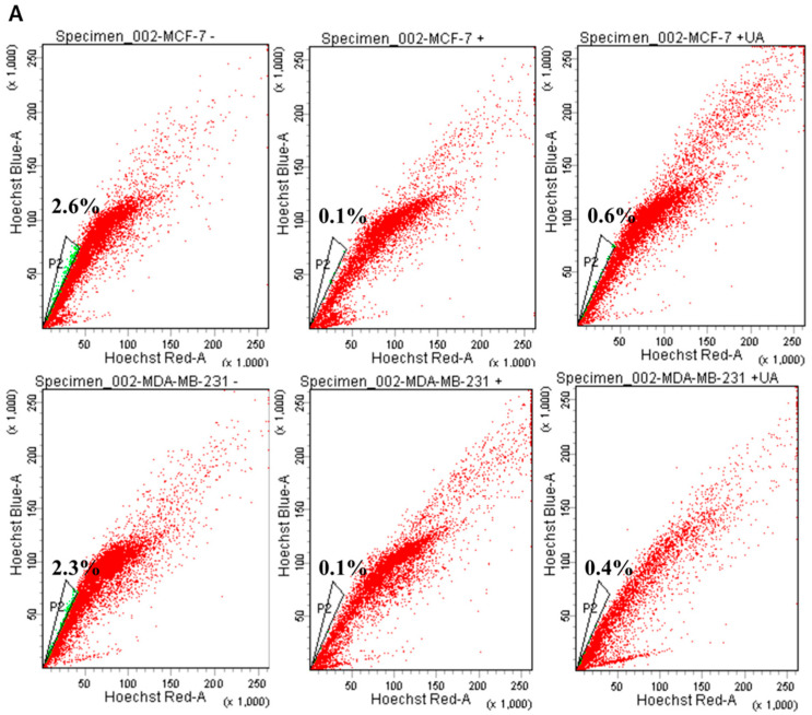
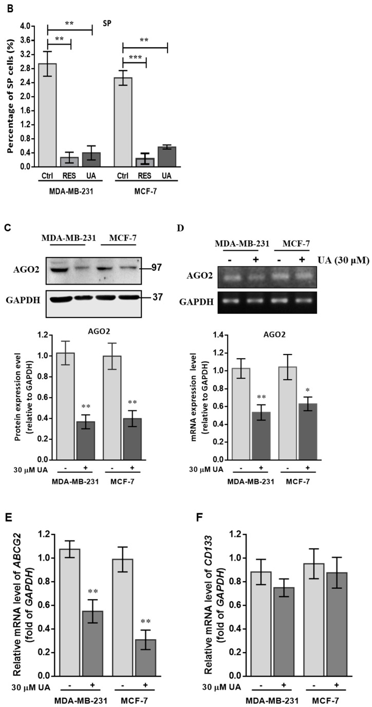
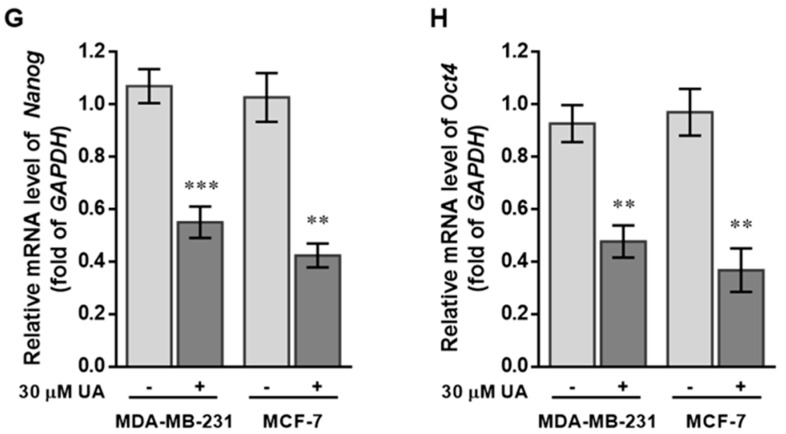
Figure 2.
UA reduces the fraction of SP in breast cancer cell lines: (A) Representative examples of cancer stem cells (SP cells) were exploited by the flow-activated cell sorting (FACS) technique. Cells were stained with Hoechst 33342 dye in the absence (left) or presence (middle) of 50 µM reserpine and 30 μM UA (right) was evaluated by FACS. The uptake of Hoechst 33342 dye occurs uniformly in all cells through passive diffusion (shown in red), SP was detected by the loss of the fluorescent population (shown in green) after treatment with the ABCG2 inhibitor (RES) or UA. (B) The relative percentage of SP was estimated in human MDA-MB-231 and MCF-7 cell lines stained with Hoechst 33342 after incubation with UA for 48 h. (C) Western blot image and (D) reverse transcription-PCR (RT-PCR) data indicate that AGO2 levels in MDA-MB-231 and MCF-7 cell lines were significantly reduced after treatment with UA relative to control cells. (E) Reduced ABCG2 associated with lower levels of CSC-related genes encoding (F) CD133, (G) Nanog, and (H) Oct4 in breast cancer cell line after UA treatment compared with the same cell line without UA treatment. GAPDH was used as an internal control for the detection of mRNA transcripts. Data were presented as mean ± SD from three independent experiments. * p < 0.05; ** p < 0.01; *** p < 0.001 vs. the untreated control.


