Figure 3.
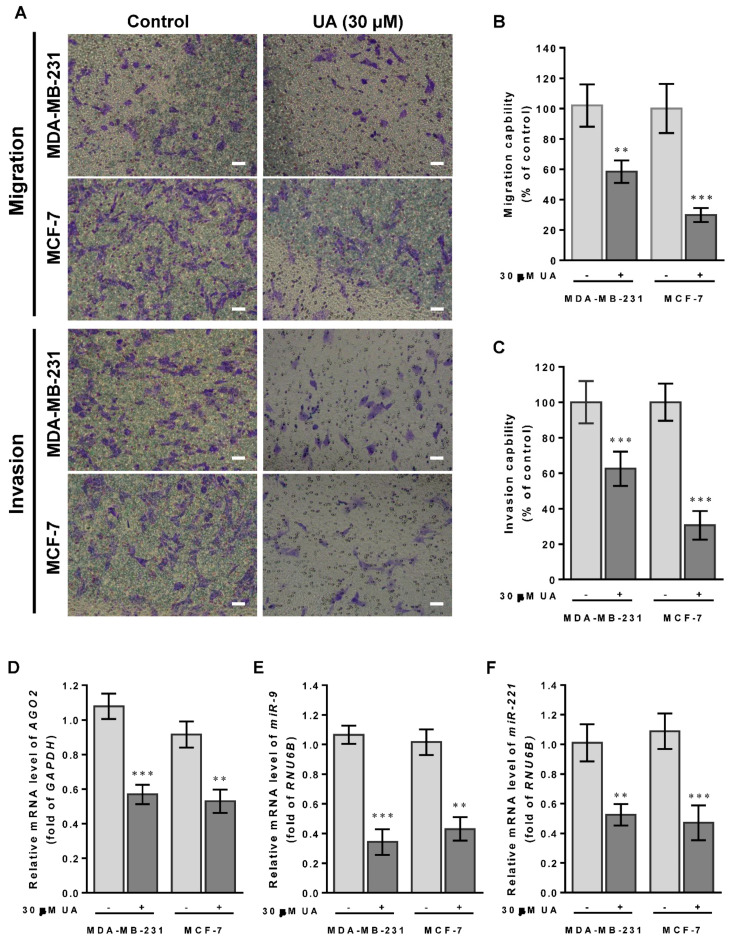
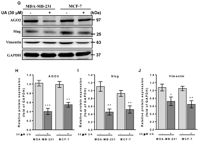
UA suppresses breast cancer cell migration and invasion: (A) Effects of UA on inhibition of cancer cell migration (top panel) and invasion (lower panel) for MDA-MB-231 and MCF-7 cells that were analyzed by the Transwell assay; scale bar = 50 μm. Quantification of migrated cells in (B) and invaded cells in (C) was presented as mean ± SD of three independent experiments. ** p < 0.01; *** p < 0.001 vs. untreated control. (D) Reduction in the AGO2 gene and EMT-related miRNA in (E) miR-9 and (F) miR-221 in breast cancer cell lines treated with UA compared with the control. Relative expression of miRNAs is quantified using TaqMan-based real-time PCR along with RNU6B. (G) Representative images of Western blot of mesenchymal proteins and densitometric analyzes of AGO2 in (H), slug in (I), and vimentin in (J) were performed using Digital Protein DNA Imagineware. GAPDH was used as an internal control. All experiments were carried out in triplicate and all data are expressed as mean ± SD. * p < 0.05; ** p < 0.01; *** p < 0.001 vs. the untreated control group.


