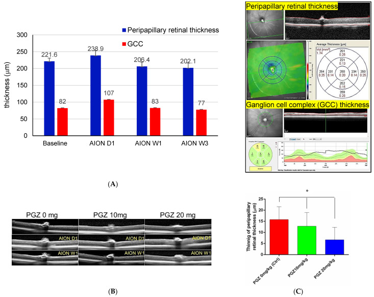
Figure 2.
OCT measurement after AION in HFD feeding followed by STZ-induced diabetic mice. (A) Peripapillary retinal thickness using posterior pole scan and ganglion cell complex (GCC) thickness were measured through OCT on day 1 (AION D1), week 1 (AION W1), and week 3 (AION W3) in diabetic mice without treatment of PGZ (n = 17). The thinning of peripapillary retinal thickness was more significant than (GCC) thickness at AION W1 and W3. The up-right figure showed how the OCT machine measured the peripapillary retinal thickness and GCC thickness. (B,C) Less retinal thinning of peripapillary retinal thickness at posterior pole scan on AION W1 was noted in DM mice treated with 20 mg/kg PGZ than in the other 2 groups (* p < 0.05).

