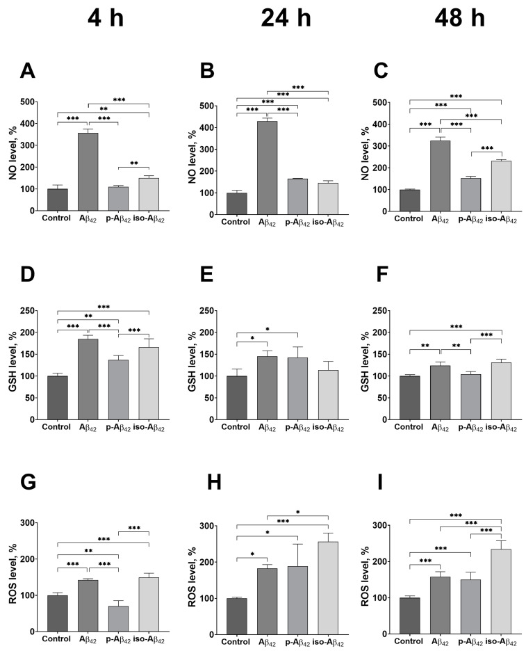
Figure 2.
The effects of beta-amyloid isoforms on the redox parameters in bEnd.3 cells. Cells were incubated for 4 h (A,D,G), 24 h (B,E,H) and 48 h (C,F,I) with 10 µM of Aβ42, p-Aβ42 and iso-Aβ42. The level of intracellular nitric oxide (NO) (A–C), reduced glutathione (GSH) (D–F) and intracellular reactive oxygen species (ROS) (G–I) were analyzed by flow cytometry. All parameters were normalized to control. The values in the control samples were taken as 100%. The mean ± SD in 3 independent experiments in triplicates is shown in the figure. The statistical difference between experimental groups was analyzed by one-way analysis of variance using the post hoc Tukey test for multiple comparisons. *—p < 0.05, **—p < 0.01 and ***—p < 0.001.

