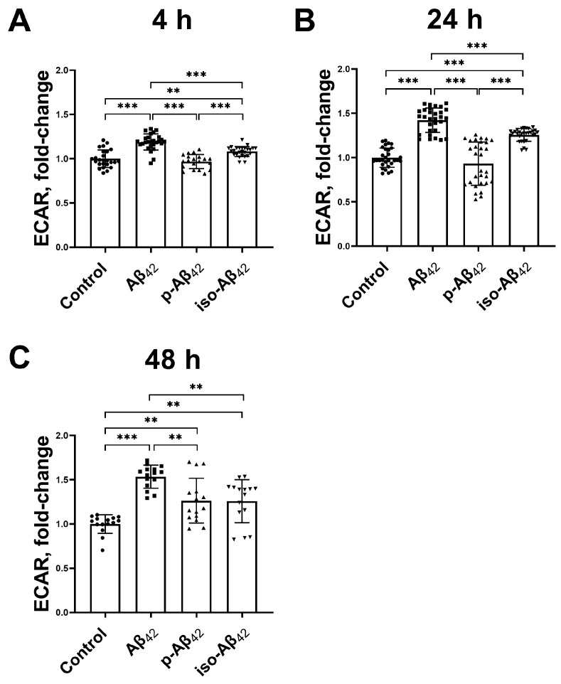
Figure 9.
Effect of beta-amyloid isoforms on the extracellular acidification rate. Mouse brain endothelial cells bEnd.3 were incubated for (A) 4 h, (B) 24 h and (C) 48 h with 10 µM of Aβ42, p-Aβ42 and iso-Aβ42. Extracellular acidification rate (ECAR)—the level of acidification of the extracellular medium. The values in the control samples were taken as 1. The histograms indicate the ratio of values in samples with beta-amyloid isoforms, normalized to the control. Each geometric figure (circle/square/up-pointing triangle/down-pointing triangle) in the histogram represents the result in an independent sample and corresponds to the Control, Aβ42, p-Aβ42 and iso-Aβ42. The mean ± SD in 3 independent experiments in 5–8 replications is shown in the figure. The statistical difference between experimental groups was analyzed by one-way analysis of variance using the post hoc Tukey test for multiple comparisons. **—p < 0.01 and ***—p < 0.001.

