Figure 1.
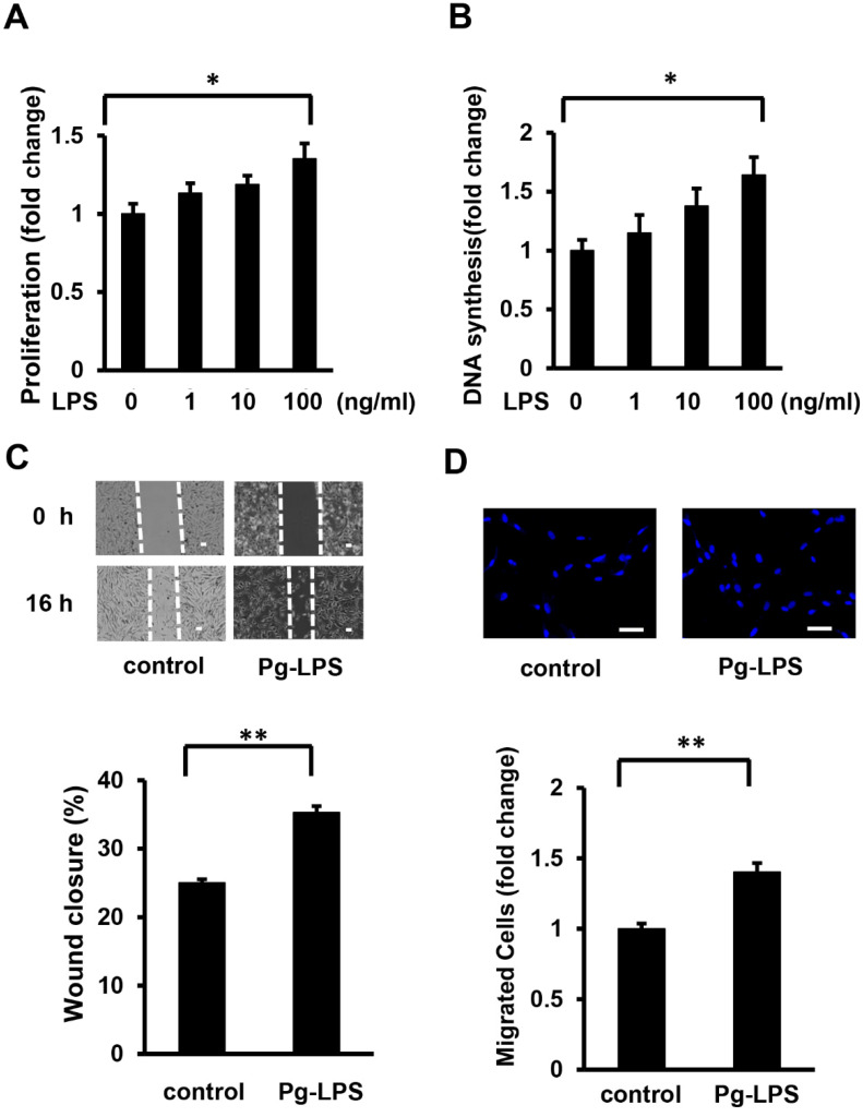
Pg-LPS induces HASMC proliferation and migration. (A) The effect of Pg-LPS on the number of HASMCs. HASMCs were treated with different concentrations (1, 10, and 100 ng/ml) of Pg-LPS for 16 h and evaluated using the MTT assay. n = 7 in each group. (B) The effect of Pg-LPS on HASMC DNA synthesis. HASMCs were treated with different concentrations (1, 10, and 100 ng/ml) of Pg-LPS for 16 h, and at the same time, cells were labeled with BrdU. n = 6 in each group. (C) The effect of Pg-LPS on HASMC migration. Upper panels show the representative scratch wound healing assay. The lower panel shows the quantitative analyses of wound closure rate. HASMCs were cultured in 6-well plates and directly scratched with a 200 μL pipette tip. Cells were incubated with or without Pg-LPS (100 ng/mL) for 16 h. n = 6 in each group. The scale bar indicates 100 μm. (D) Effect of Pg-LPS on the migratory property of HASMCs. The upper panels show the representative DAPI staining of migrated cells. HASMCs were seeded in a transwell and treated with or without Pg-LPS (100 ng/mL) for 16 h. n = 4 in each group. The scale bar indicates 50 μm. * p < 0.05 vs. basal; ** p < 0.01 vs. basal. Data are presented as mean ± SE.

