Figure 4.
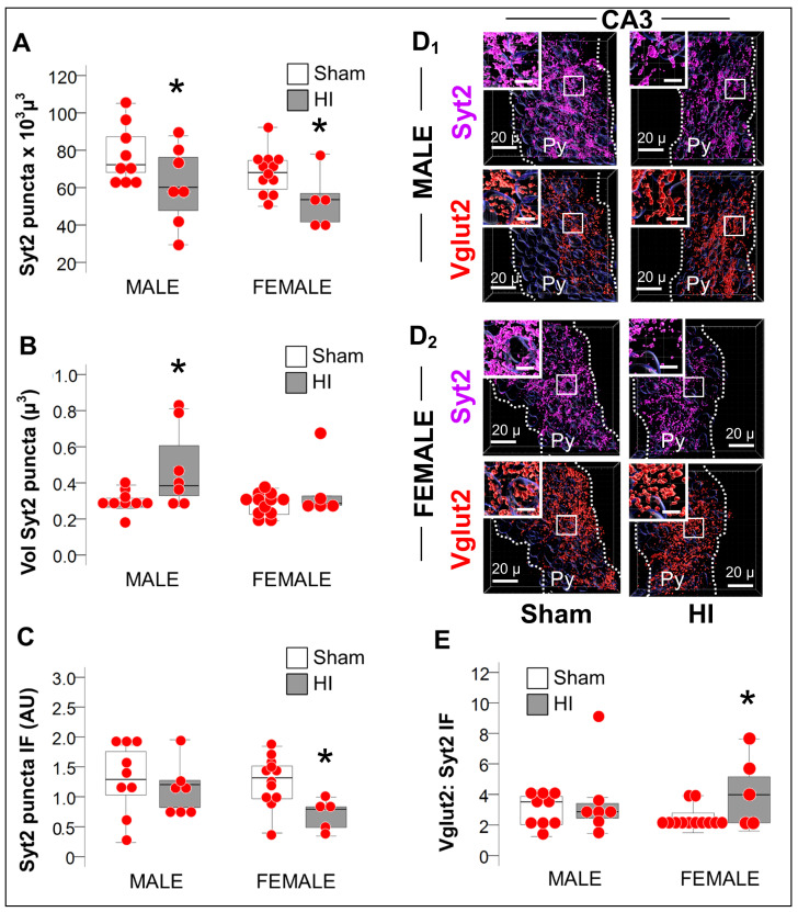
Decreased Syt2 puncta and predominance of excitation in female mice after HI. (A) The number of the puncta expressing the PV-specific presynaptic marker Syt2 was similarly decreased in the CA3 of both males and females HI-injured mice (grey box) compared to sham (white box). (B) Unlike, in female mice, CA3 Syt2+ puncta from HI-injured male mice (grey box) were larger in volume compared to sham (white box). (C) Syt2 immunofluorescence per puncta in arbitrary units (AU) was decreased in HI-injured female mice (grey box) compared to sham (white box), but not in male mice. (D) Representative Imaris reconstructions of Syt2 (magenta) and the glutamatergic Vglut2 (red) presynaptic markers within the pyramidal cell layer (Py) for male (D1) and female (D2) sham and HI-injured mice are shown. Radiatum (Rd) and Oriens (Or) layers were negated, to only account for perisomatic synapses within the Py. (E) The Vglut2:Syt2 ratio was greater in the HI-injured CA3 of female mice (grey box) supporting the EcFP LTP results shown in Figure 2D. Quantifications are shown as hybrid box, whisker and dot plots. *, p <0.05 (Mann-Whitney U test; n= 6, per sex per group).

