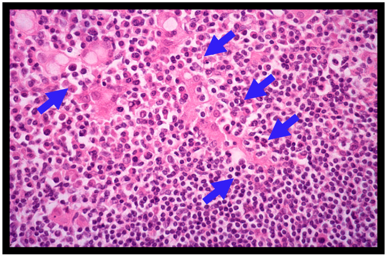
Figure 2.
Histological features of gastric MALT lymphoma (H&E; X400). Diffuse infiltrate of small- to medium-sized atypical neoplastic lymphoid cells (centrocyte-like cells) around reactive follicles with a marginal zone growth pattern is shown. Lymphoepithelial lesions (LELs) (blue arrows) are also shown.

